
Digital X-Rays in Darien
Digital radiographs that reveal decay, bone loss and cracks a visual exam cannot see: on screen instantly, with no darkroom and less radiation.
Most of a tooth is not visible when you open your mouth. X-rays are how the parts that matter — between the teeth, below the gum, inside the root — get examined at all.
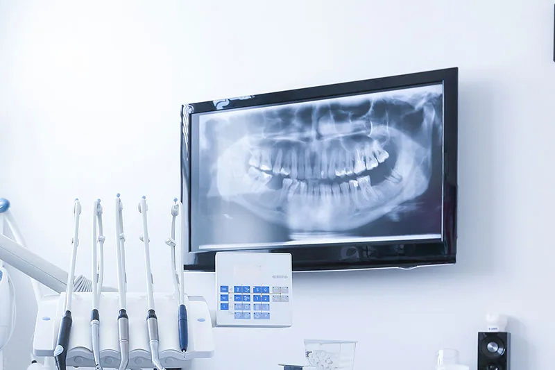
What’s included
- Detects decay between teeth and bone loss
- Image appears on screen immediately — no darkroom
- Essential for planning root canals and implants
- Images compared digitally against your previous visits
- Finds cracks in fillings invisible to the naked eye
About Digital X-Rays
Dental X-ray images, also known as dental radiographs, are one of the most important pieces of equipment a dentist has for keeping teeth, gums and mouth healthy. A dental X-ray allows dentists to detect problems that are just not visible to the naked eye, such as cracks in fillings, decay in between teeth and bone loss. Dental X-rays are also vital when planning dental work such as root canals and dental implants. The main differences between digital dental X-rays and regular X-rays are their ease of use. In digital radiography, the film is replaced with a digital dental x-ray sensor. This digital dental X-ray sensor receives the X-rays in much the same way as a film does, but instead of developing the film in a dark room, the digital dental X-ray sensor sends the image directly to a computer where it appears on the screen. As well as the speed and convenience of this process, one of the major advantages of digital dental X-rays is that a computer can digitally compare an image to previous images.
What X-Rays Find
Decay between teeth, where the surfaces touch and neither a mirror nor a probe can reach. This is where a large proportion of cavities start and where they are invisible until they are large. Decay underneath existing fillings and crowns. Cracks and breakdown in old restorations. Bone loss from periodontal disease, which is how gum disease is actually staged. Infection or an abscess at the root tip, often before there is any pain. Cysts and other changes in the jaw. Impacted teeth and wisdom teeth that have not come through. The position of roots relative to the sinus and the nerve, which is essential before extractions and implants. And teeth still developing in children, so problems with eruption are caught while there is time to guide them.
Benefits of Digital Radiography
Digital sensors need substantially less radiation than the film they replaced, which is the benefit most patients ask about first. The image appears on the screen within seconds rather than needing chemical development, so we can discuss it with you while you are still in the chair, and if the angle was not quite right, retake it immediately instead of at a second appointment. On screen, images can be enlarged, contrast-adjusted and measured, which makes early decay easier to identify than on a small film held up to a light. And because everything is stored, a computer can digitally compare an image to previous images, so a shadow that has grown slightly since your last visit becomes obvious in a way that would be very difficult to judge from memory.
What to Expect
A small sensor is placed inside your mouth and you bite gently to hold it in position while the image is taken; each one takes a couple of seconds and there is nothing to feel. A lead apron with a thyroid collar is used. How many and how often depends on your risk. Someone with a history of decay or gum disease needs bitewings more frequently than someone with a stable mouth, and X-rays are taken because they will change what we do, not on a fixed schedule regardless. Afterward we will put the images on the screen and show you what we are looking at. If you are pregnant or think you might be, tell us, and we will postpone anything non-urgent.
Common Questions
How much radiation is involved?
How often do I need them?
Can I refuse X-rays?
Can I get X-rays while pregnant?
Can you use X-rays from my previous dentist?
Terms You May Hear
- Bitewing
- The most common X-ray, showing where upper and lower back teeth touch: the classic site for hidden decay.
- Periapical
- An image of a whole tooth including the root tip and surrounding bone, used to check for infection.
- Radiograph
- The clinical name for an X-ray image.
Related Services
Ready to Schedule Your Appointment?
Call (630) 427-1800 to book an exam. If you have recent X-rays from another office, we can request them.